![]()
![]()
![]()
5.1 Introduction
5.2. Isolating infective larvae from herbage
5.3 Packed cell volume determination (PCV, haematocrit)
This chapter considers two supplementary diagnostic procedures. The first is the isolation and identification of infective larvae from the herbage. This procedure allows the estimation of larval availability on the pasture and can be used to define larval seasonality and distribution. The second is the estimation of anaemia in clinically or sub-clinically affected animals by the determination of packed cell volume (PCV) in a blood sample.
5.2.1 Principle
5.2.2 Application
5.2.3 Equipment
5.2.4 Procedure
This is a qualitative and semi-quantitative procedure for isolating, counting and identifying L3 from representative samples of herbage taken from defined grazing areas.
This is a useful procedure for determining seasonal variations in L3 availability on the pasture. However, the number of larvae on the herbage is influenced by a variety of factors, and may vary considerably even when repeated samplings are done at the same time of day and the same site; herbage larval counts should therefore be interpreted with extreme caution.
· Scissors and collecting bags· Small bucket (e.g., 7 litre), calibrated at one-half litre gradations
· Gauze bag, made from terylene netting or cheesecloth, large enough to fit over the rim of the bucket
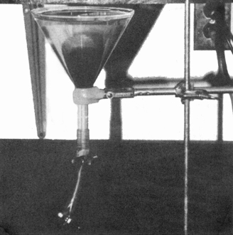
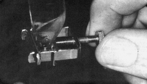
· Large plastic or glass filter funnel (20 cm diameter), fitted with a length of flexible, transparent tubing carrying 2 screw clamps arranged so that about 15 ml can be trapped between the two clamps
· Domestic detergent
· Test tube, holding at least 15 ml
· Dissecting microscope or microscope
· Balance
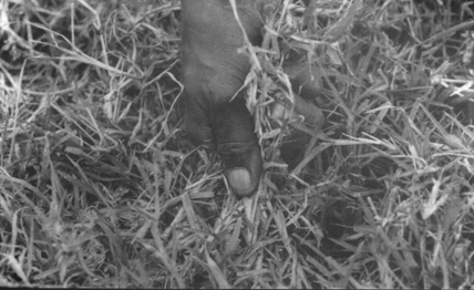
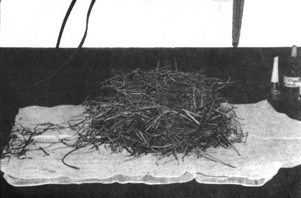
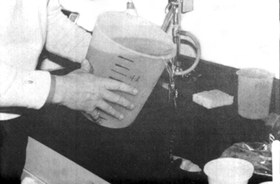
(a) Collect small samples of grass from a large number of sites randomly scattered throughout the area to be sampled, using a "W" or "N" collecting route. Avoid areas of heavy faecal contamination and avoid collecting soil.
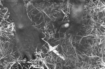
(b) Grass can be collected by hand. Where grasses are coarse, scissors may
be used. Grass samples of approximately 300-600 g should then be placed in a
plastic bag.
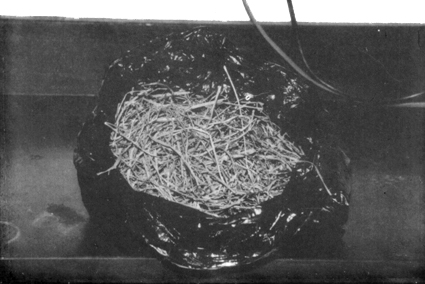
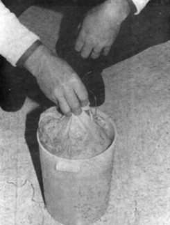
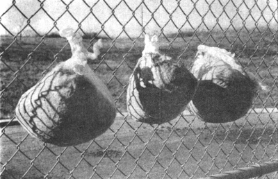
(c) Place the collected grass sample inside a gauze bag and immerse the bag in water in a bucket but keep the bag clear of the bottom of the bucket.
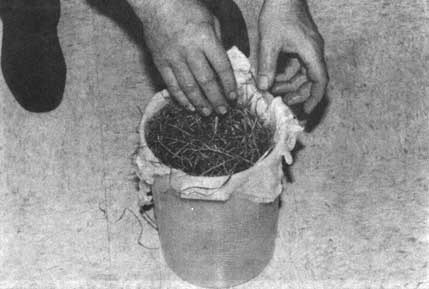
(d) In the first 3-4 hours, remove, drain and replace the bag in the water several times to agitate the sample. Leave the bag in the water at room temperature overnight.
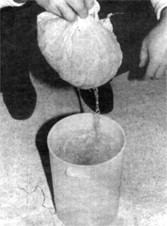
(f) The bag of grass should be dried (sun/oven/incubator) and weighed when completely dry.
(i) Leave the funnel to stand for another hour.
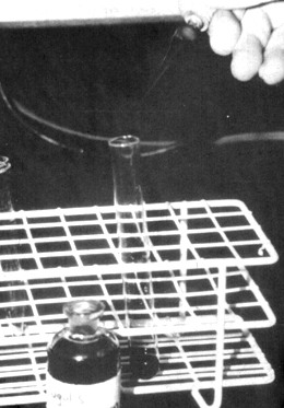
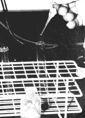
(k) Siphon off the supernatant to leave 35 ml, which is stained with 35 drops of iodine for at least 1 hour. Counter stain with 3 drops of sodium thiosulphate.
(l) Count and identify the parasitic larvae seen. There will usually be many more non-parasitic nematodes and larvae present than parasitic larvae and the count may therefore be rather laborious.
(m) Parasitic larvae all have sheaths discernible at the tail end and tend to retain the brown coloration of the iodine for 30-40 minutes after decolouration with sodium thiosulphate.
Larval identification keys can be found in Tables 3.3 and 3.4.
(n) Number of larvae per kg of dry herbage = count x 1,000/weight of dry herbage (in grams).
5.3.1 Principle
5.3.2 Application
5.3.3 Equipment
5.3.4 Procedure
Infections with some parasite species, particularly Haemonchus but also Bunostomum, Trichuris and Fasciola can cause anaemia. In acute haemonchosis and fascioliasis, the pathogenic effect of the parasite is often present before eggs appear in the faeces. The PCV technique allows an estimation of the degree of anaemia present by measuring the volume occupied by the red blood cell in a sample of circulating blood.
The PCV determination is a useful procedure to carry out on both individual animals and herds/flocks to assess the possible role of Haemonchus and other blood-sucking parasites in a parasite problem. It is useful as an early aid to the diagnosis of haemonchosis and fascioliasis. However, anaemia can occur as a result of other causes, in particular trypanosomiasis and some tick-borne diseases, so this test must be done in conjunction with both parasite egg counts and assays for circulating haemoparasites.
· Needles and syringe (or Vacutainer needles and tubes containing anticoagulant)
· Micro-haematocrit tubes
· Micro-haematocrit centrifuge
· Micro-haematocrit reader
· Haematocrit tube sealant
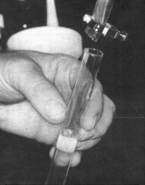
(a) Take a venous (jugular or tail) blood sample into a test tube or Vacutainer containing anticoagulant (such as EDTA).(b) Mix the blood sample well but gently for 2 minutes.
(c) Draw the well-mixed blood up a 75 x 1.5 mm capillary tube for ¾ of its length.
(d) Seal one end with sealant.
(e) Place in the micro-haematocrit centrifuge, ensuring that the sealant is at the outer end.
(f) Close the centrifuge lid.
(g) Centrifuge the tubes at 12,000 rpm for 4 minutes.
(h) Place the tubes in the reader and note the reading.
(i) Express the reading as a percentage of packed red cells in the total volume of whole blood.
![]()
![]()
![]()